- 2026年6月7日
[Explained by a Specialist] What Is Your Fatty Liver Level? How to Determine Severity Through an Ultrasound Examination
“I was told I have a fatty liver at my health checkup, but I don’t have any particular symptoms…”
“My liver enzyme levels seem a little high, but I guess I’m fine?”
Is there anyone who thinks this way? The liver is known as a “silent organ.” Precisely because it rarely shows symptoms, it is extremely important to accurately understand the current condition of your liver.
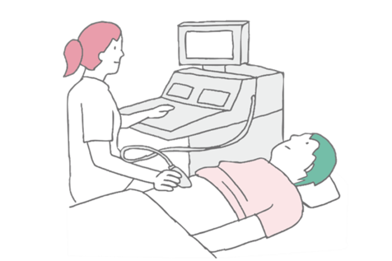
Even though we simply call it “fatty liver,” there are actually differences in its “degree” or severity. To understand this degree, an “ultrasound (echo) examination” is incredibly useful.
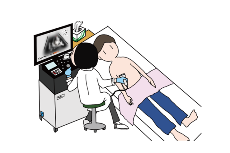
At Saito Clinic of Internal Medicine,, we place great emphasis on ultrasound examinations when evaluating liver health. This is because it is an excellent examination method that provides a wealth of information about the liver without placing any physical burden on the patient.
In this article, we will explain in detail from a specialist’s perspective how a fatty liver looks on an ultrasound, how severities such as “mild,” “moderate,” and “severe” are judged, and why knowing this is so important.
What Exactly Is a Fatty Liver? Why Can It Be “Seen” on an Ultrasound?
Fatty liver, as the name suggests, refers to a condition where an excessive amount of neutral fat (triglycerides) has accumulated inside the liver cells. Causes include not only alcohol consumption but also overeating, a lack of exercise, obesity, and diabetes.
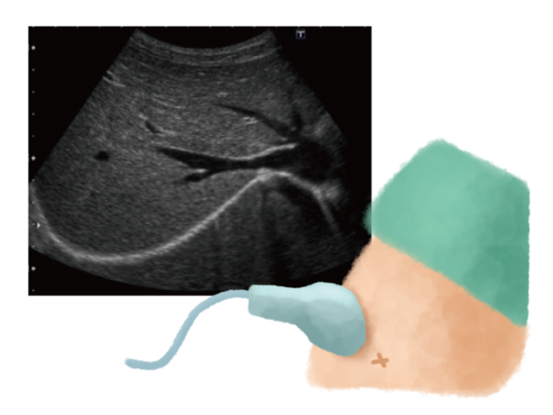
So, why can the fat accumulated in the liver be “seen” on an ultrasound?
An ultrasound examination uses high-frequency sound waves (inaudible to the human ear) applied from the body’s surface, capturing the reflected waves (echoes) bouncing back from your organs to display them as an image. It works on the same principle as a fish finder.
When a lot of fat accumulates in the liver, the ultrasound waves reflect more strongly than usual. As a result, on the ultrasound image, it appears “whitish (technically known as hyperechoic)” compared to a healthy liver. We evaluate the severity of the fatty liver based on the degree of this “whiteness” and differences in its appearance.
Incidentally, an ultrasound is an extremely safe examination with no pain and no concerns about radiation exposure.
Severity of Fatty Liver on an Ultrasound: Distinguishing Between “Mild,” “Moderate,” and “Severe”
During an ultrasound examination, we mainly evaluate the severity of a fatty liver in three stages—”mild,” “moderate,” and “severe”—based on the following points:
- Whiteness of the liver (Hepatorenal contrast): We compare the appearance of the liver with the adjacent kidney. The more fat there is, the whiter the liver looks.
- Appearance of blood vessels within the liver: The walls of the blood vessels (such as the portal vein) running through the liver become harder to see due to fat accumulation.
- Appearance of the deep parts of the liver (Deep attenuation): If there is a lot of fat, it becomes difficult for the ultrasound waves to reach deep into the liver, making the deeper areas unclear.
- Appearance of the diaphragm: The outline of the diaphragm located above the liver becomes difficult to see.
By comprehensively evaluating these findings, we classify the condition as follows:
[Mild Fatty Liver]
- The entire liver looks slightly whitish compared to the kidney.
- The walls of the blood vessels inside the liver can still be seen relatively clearly.
- The deeper parts of the liver and the diaphragm can also be observed without issues.
[Moderate Fatty Liver]
- The whiteness of the liver increases further, and the color difference from the kidney becomes more distinct.
- The walls of the blood vessels inside the liver become harder to see.
- The deeper parts of the liver may appear slightly blurry (deep attenuation).
[Severe Fatty Liver]
- The entire liver appears extremely white and bright.
- The walls of the blood vessels inside the liver become almost invisible.
- The deeper parts of the liver and the diaphragm become quite unclear.
Why Is It Important to Know the “Severity”?
You might wonder, “What difference does it make knowing the degree of my fatty liver?” However, knowing the severity evaluated by an ultrasound is extremely important for your future health management.
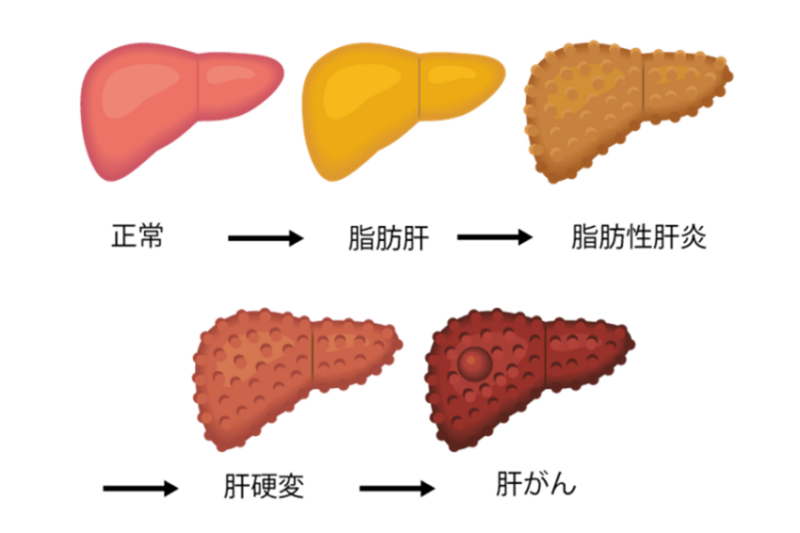
- Indicator of progression risk: If left untreated, a fatty liver risks progressing to inflammatory conditions such as “Non-Alcoholic Steatohepatitis (NASH)” and “Alcoholic Hepatitis,” and further advancing to “liver cirrhosis” (where the liver hardens) and “liver cancer.” A higher severity on the ultrasound (especially moderate to severe) suggests a higher potential risk of such progression.
- Possibility of fibrosis (hardening of the liver): If the degree of fat shown on the ultrasound is severe, or if the edges of the liver appear rounded or the surface looks bumpy, it is possible that fibrosis (hardening) of the liver is advancing. (Note: It is difficult to accurately diagnose fibrosis using only an ultrasound, and other tests may be necessary.)
- The necessity for countermeasures changes: For mild fatty liver, the primary focus will be strictly on lifestyle improvements. However, for moderate or higher severity, or if progression is suspected, stricter management, detailed evaluations via blood tests (fibrosis markers, etc.), specialized tests to measure liver stiffness (such as Shear wave elastography), and in some cases, medical treatment may be required.
In short, grasping the “level” of your fatty liver through an ultrasound is the first step toward taking appropriate measures and preventing serious diseases in the future.
Why Our Clinic Focuses on “Ultrasound Examinations”
At our clinic, we position the ultrasound (echo) examination as an extremely vital test to understand the liver’s condition. The reasons for this are as follows:
- Low burden on the patient: There is no pain during the examination, allowing you to undergo it in a relaxed state. The examination time is also relatively short.
- High safety: Since no radiation is used, there are no concerns about radiation exposure, making it safe for pregnant women or those who require repeated testing.
- Real-time observation: We can conduct detailed evaluations because the examination is performed while observing the inside of the liver in real time on a monitor. While CT scans leave slight gaps between slices that might miss certain areas, ultrasounds allow for continuous, seamless observation.
- Detection of lesions other than fatty liver: It also serves as an opportunity to discover various abdominal diseases—not just the degree of fat, but also abnormalities in the shape and size of the liver, liver tumors (such as cancer), gallstones, gallbladder polyps, and abnormalities in the pancreas or blood vessels.
- High-quality diagnosis by a specialist: At our clinic, the director—who is a specialist in gastroenterology, hepatology, and ultrasound—carefully performs the examinations with the utmost attention. The quality of a diagnosis is greatly influenced not only by the equipment but also by the skill and experience of the performing physician. We always strive for high-quality examinations so as not to miss even the slightest changes.
The Limits of Ultrasound Examinations and the Next Steps
While ultrasound examinations are excellent for observing the degree of a fatty liver and the morphology of the liver, they are not perfect. For example, there are limits to accurately diagnosing whether the fatty liver is accompanied by inflammation (whether it is NASH) or to what extent liver fibrosis (hardening) has progressed.
Therefore, if moderate or higher fatty liver is found on an ultrasound, or if there are findings suggesting progression to liver cirrhosis, the following steps may be necessary:
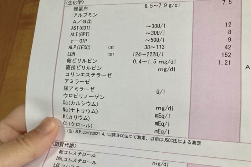
- Blood tests: In addition to re-evaluating liver function (AST, ALT, γ-GTP, etc.), we check the platelet count and markers that estimate the degree of fibrosis (such as the FIB-4 index).
- Measurement of liver stiffness (Elastography): At our clinic, we use applied ultrasound technologies like Shear wave to numerically evaluate the stiffness of the liver. The degree of fibrosis can be estimated without performing a liver biopsy.
- Liver biopsy: If necessary, this is an examination where a thin needle is inserted into the liver to collect a small tissue sample, which is examined in detail under a microscope. It can diagnose the degree of inflammation and fibrosis most accurately.
The ultrasound examination serves as a crucial gateway to determine whether these more detailed tests are needed.
Knowing Your Liver’s Condition Through Ultrasound for Future Health

Fatty liver is no longer a disease exclusively for “people who are overweight” or “people who drink alcohol.” Furthermore, it is not a disease where you can rest easy just because “there are no symptoms.”
An ultrasound (echo) examination is a highly useful tool that can safely and painlessly evaluate how much fat has accumulated in your liver and its “severity.” Knowing your stage of progression—mild, moderate, or severe—is an important step in properly understanding your health condition and starting appropriate countermeasures.
At our clinic, the director, who is a specialist in gastroenterology, hepatology, and ultrasound, responsibly performs the ultrasound examinations and carefully evaluates your liver’s condition. If you have been told you have a fatty liver at a health checkup, are concerned about your liver values, or want to thoroughly check your liver’s condition, please feel free to consult us.
And even if your liver condition has progressed, please do not give up. At our clinic, we are also engaged in regenerative liver medicine as a new treatment option for advanced liver diseases. Let’s start by knowing the “now” of your liver through an ultrasound examination. That will be the first step toward your future health.
In this article, we explained how to determine the severity of a fatty liver using an ultrasound examination. For those suffering from daily symptoms and their families, we understand that encountering this information may bring about various feelings, including anxiety and hope for future treatments.
At Saito Clinic of Internal Medicine, we are focusing on a new approach called “regenerative liver medicine” for patients facing the progression of such liver diseases.
“Is there still something that can be done for my condition?”
“What exactly is regenerative medicine?”
You may have such questions or hopes.
Our clinic director has stood at the forefront of liver disease treatment for many years. We have prepared a video in which the director directly and clearly explains “About Our Clinic’s Regenerative Liver Medicine” and the possibilities it brings.
▼ Click here for the explanation video on regenerative liver medicine by our director
By watching this video, we hope you will deepen your understanding of what regenerative medicine entails and the dedication with which our clinic approaches this treatment.
If you watch the video and find yourself even slightly interested, please do not struggle alone; feel free to consult with us. You can contact us by phone or through the inquiry form on our clinic’s website.
We also offer preliminary consultations via online telemedicine, so please feel free to reach out.
Let’s nurture hope for your future together.
Information on Online Preliminary Consultations

さいとう内科クリニック
院長:斉藤 雅也 Masaya Saito
- 日本内科学会認定医
- 日本肝臓学会専門医
- 日本消化器病学会専門医
- 日本超音波医学会専門医
- 日本消化器内視鏡学会専門医

神戸大学医学部附属病院等の最前線で長年消化器・肝臓内科の臨床と研究に従事。医学博士。 標準治療では回復が困難な進行した肝炎や肝硬変に対し、新たな選択肢としての「肝臓再生医療」にいち早く取り組む。また、肝硬変患者さまの中で合併症(潜在性肝性脳症)を有する割合を明らかにし、カルニチンによる潜在性肝性脳症の治療効果を世界で初めて報告するなど、国際的な英文医学誌への論文掲載実績も多数(代表論文:Hepatol Res 2016; 46(2): 215-224)。科学的根拠に基づいた高度な専門知識と精緻な診断で、患者様の肝臓を守るサポートを行っています。
≫ 詳しい経歴や全研究実績はこちら
- 院長
- 斉藤雅也 Masaya Saito
日本肝臓学会 肝臓病専門医 Hepatologist, The Japan Society of Hepatology - 所在地
- 〒651-2412
兵庫県神戸市西区竜が岡1-15-3
(駐車場18台あり) - 電話
-
- 電話:078-967-0019
- 携帯電話:080-7097-5109
- アクセス
- 当院は、神戸市西区と明石市の境界付近に位置しており、明石市からも徒歩圏内です。実際に、明石市方面からも多くの患者様(肝臓病・一般内科)にご来院いただいております。駐車場も完備しておりますので、お車での通院も便利です。